Submitted on December 16, 2020

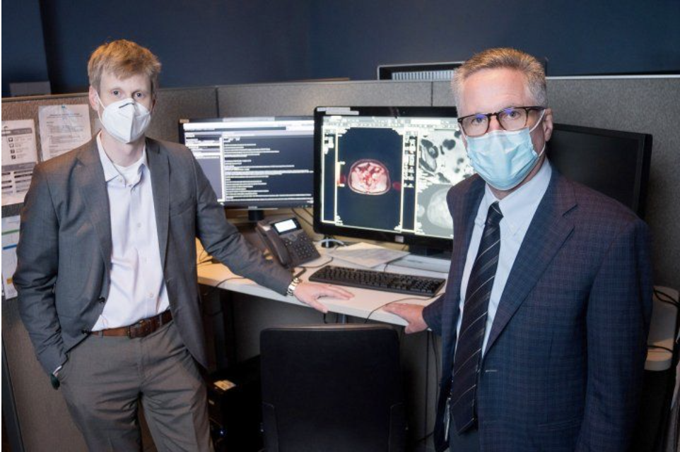
FDA approval right out of Academia doesn’t happen often nevertheless for these collaborators, UCSF and UCLA, together they accomplished this. At both institutions, faculty from the Departments of Radiology, Urology, Radiation Oncology and Medical Oncology led this endeavor. Study investigators included UCSF’s Dr. Thomas Hope, Director of Molecular Therapy in the Department of Radiology and UCLA’s Drs. Johannes Czernin, Chief and Jeremie Calais, Assistant Professor, both at the Ahmanson Translational Imaging Division of the Department of Molecular and Medical Pharmacology. To support these studies UCSF Urology through philanthropy supplied the resources necessary for the study at UCSF. Among these philanthropists include the Prostate Cancer Foundation, who has invested over $26 million in research on PSMA since inception in 1993.
These pivotal studies, two prospective multi-site clinical trials were performed in which, 960 men with prostate cancer underwent the PSMA PET technique. At UCSF the Department of Urology, drove a strong and very substantial recruitment effort. In one study investigators evaluated the PET imaging agent (Ga- 68-PSMA-11) for its ability to detect prostate cancer cells in patients with biochemical recurrence after prostatectomy and radiation therapy. In another study, investigators aimed to see how well this PET imaging agent can detect prostate cancer that has recurred in patients after initial therapy. The findings in these studies project that PSMA PET technique can detect the location prostate cancer cells earlier and more effectively than current imaging techniques. Learn more about these studies at ClinicalTrials.org: Gallium-68 PSMA-11 Positron Emission Tomography (PET) Imaging in Patients With Biochemical Recurrence and Gallium Ga 68-labeled PSMA-11 PET/CT in Detecting Recurrent Prostate Cancer in Patients After Initial Therapy
Patients may be eligible for PSMA PET imaging at initial diagnosis with prostate cancer or who have been already diagnosed, treated and their prostate cancer returned. In this imaging technique, the PET imaging agent is injected into the body and attaches to proteins known as prostate-specific membrane antigens. Prostate cancer cells overexpress these proteins on their surface. This agent (GA-68-PSMA-11) a radioactive tracer drug highlights, in the imaging scan the places where the prostate cancer cells are located.
Information about PSMA PET patient care, can be found at UCLA and UCSF websites.
“I believe PSMA PET imaging in men with prostate cancer is a game changer because its use will lead to better, more efficient and precise care,” said Peter Carroll, MD, MPH, a professor at the UCSF Helen Diller Family Comprehensive Cancer Center.
Dr. Carroll is one of the UCSF Department of Urology’s Faculty who has played an integral role in prostate research including its induction, detection, and treatment. Furthermore, members of the UCSF Department of Urology, including Drs. Carroll and Nguyen, have been involved in additional trials (OSPREY and CONDOR), of products such as PyLTM, an investigational fluorinated PSMA-targeted PET imaging agent that allows one to see localized prostate cancer, in addition to bone and soft tissue metastases to help detect recurrent metastatic prostate cancer, which recently granted priority review and is next in line for FDA approval. They are also, leading a phase one trial with Intuitive Surgicals Inc. for an intraoperative PSMA fluorophore which is designed to enhance direct visualization and removal of microscopic cancerous tissues outside the prostate and nearby lymph metastatic lymph nodes during robotic surgery. This innovative study is first in human and only open at UCSF. All of these trials are a reflection of the Department’s deep commitment to the improved care of men with prostate cancer. The department is deeply committed to collaboration which enables such discoveries and is very grateful to those advocates who supply the resources to test new technology.