Submitted on December 7, 2015

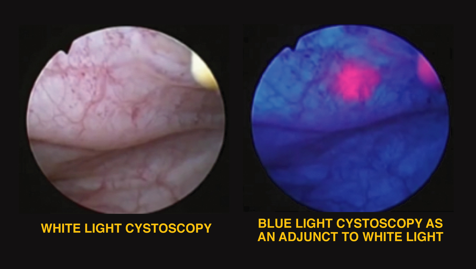
Correctly imaging a tumor is a key element in successful surgical treatment of bladder cancer. Typically, a cystoscope is introduced through the urethra into the bladder, and suspicious lesions are visualized with white light emitted by the scope. The lesions are then removed by introducing additional instruments into the cystoscope in a procedure called transurethral resection.
To improve detection of tumors, UCSF Urology has begun using a new imaging system that exploits the fluorescent properties of naturally occurring molecules in malignant tissues. An hour before the cystoscopy, an imaging solution is delivered into the bladder, which is preferentially absorbed by cancerous tissue. The doctor then inserts a cystocope that has the ability to emit blue light as well as white light. The blue light reveals cells that have absorbed the contrast solution, allowing physicians to detect tumors that are not visible with white light alone.
Kirsten Greene, MD, MS, Associate Professor of Urology at UCSF and San Francisco Veterans Affairs Medical Center (SFVA), has been using the system at for ## months at the VA, with excellent results. The imaging option is now available at UCSF Mission Bay as well, where Sima Porten, MD, MPH, directs its use.
For more information, contact 415.353.7171
